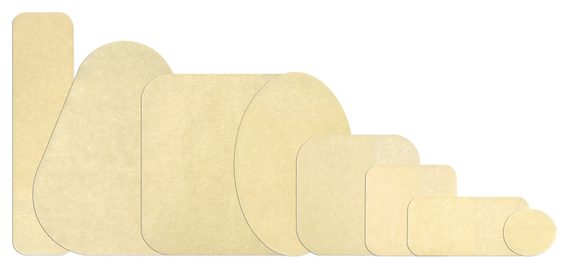
DuoDerm® Extra Thin Dressing
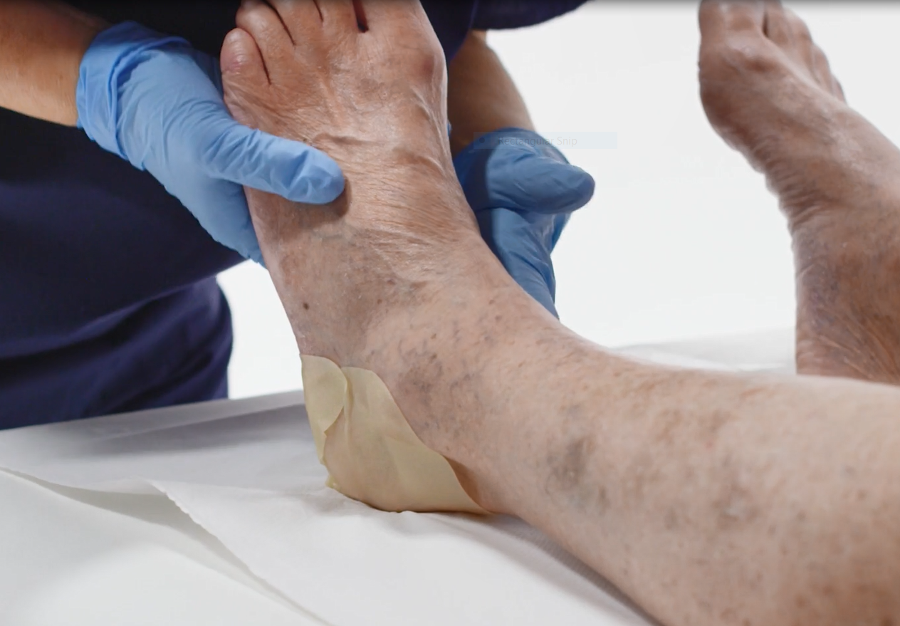
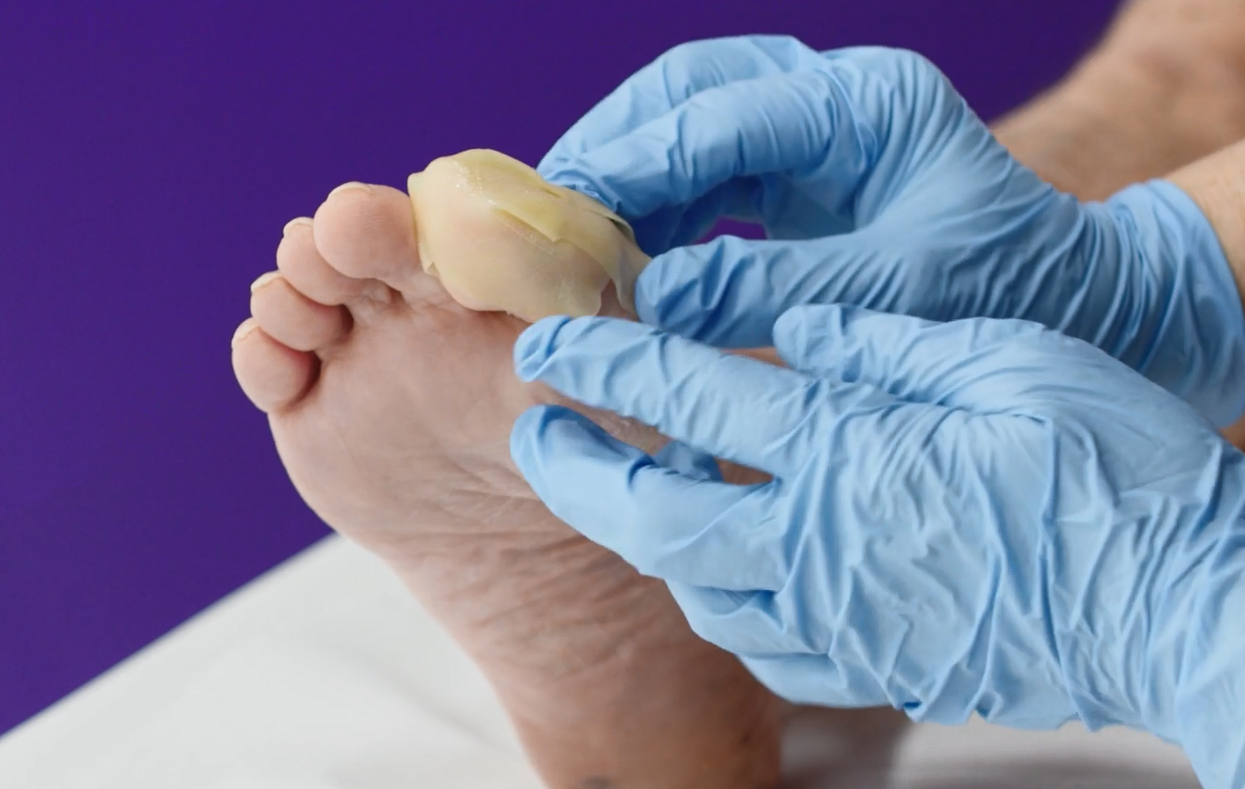
Designed to be used as a primary or secondary dressing for the management of wounds and for the protection of intact skin. DuoDerm® Extra Thin dressings are easy to use¹ and can be cut to shape¹ to allow the convenient management of wounds in awkward areas.
 ;
;
Not all hydrocolloids are the same
Hydrocolloid dressings consist of a gel-forming agent such as carboxymethylcellulose or pectin, which interacts with wound exudate to create a moist interface that promotes autolytic debridement and minimises pain during dressing changes. DuoDERM™ dressings provide a moist wound environment to promote autolytic debridement², creating an occlusive, moist wound environment that supports reepithelialisation.³
Our family of products is designed to bring gentleness to wound management, to give patients peace of mind and to make your life easier as hydrocolloid technology does not restrict joint motion or patient mobility⁴.
 ;
;
Creating a moist wound-healing environment that supports re-epithelialization
DuoDerm™ Extra Thin dressings are easy to use and can protect against the spread of viruses such as HBV and HIV-1*⁵ and the spread of bacteria including MRSA.**⁶
Nothing grabs your attention like a wound "on the edge", and this may or may not make the transition for wound management into an unfavorable situation. Wound dressings manage this situation by being applied at the right time to increase the chances of successful healing. Duoderm ™ Extra Thin promotes a faster rate of healing compared to traditional gauze dressings⁷ and they may increase the likelihood of pressure ulcer healing compared with other hydrocolloids⁸.
 ;
;
DuoDerm® Extra Thin use in wound care
DuoDerm® Extra Thin dressings are indicated for:
- Pressure injury/ulcers
- Surgical wounds
- Partial thickness burns
- Abrasions/lacerations
The European Pressure Ulcer Advisory Panel (EPUAP) and The National Pressure Ulcer Advisory Panel (NPUAP) guidelines recommend the usage of hydrocolloids for the management of pressure ulcers.⁹
DuoDerm® Extra Thin dressings provide a physical, microbial, viral, and waterproof barrier to protect the wound.
*In in-vitro testing.
**Whilst the dressing is intact and without leakage.
1. Forshaw A. Hydrocolloid dressings in paediatric wound care. J Wound Care 1993; 2(4): 209–12.
2. Pudner R. Hydrocolloid dressings. PN 1998; 9: 7–19.
3. Greguric S, Budimcic D, Soldo BA et al. Hydrocolloid dressing versus a conventional dressing using magnesium sulphate paste in the management of venous leg ulcers. Acta Dermatovenerol Croat 1994; 2: 65 71.
4. Hopper 2012 Enhancing patient recovery following lower limb arthroplasty with a modern wound dressing: a prospective, comparative audit
5. Bowler PG, Delargy H, Prince D and Fondberg L. The viral barrier properties of some occlusive dressings and their role in infection control. Wounds 1993; 5(1): 1–8.
6. Wilson P, Burroughs D and Dunn L. Methicillin resistant staphylococcus aureus and hydrocolloid dressings. Pharm J 1988; 241(1): 787–88.
7. Demetriades D and Psaras G. Occlusive versussemi-open dressings in the management of graft-donor sites. South African Journal of Surgery.1992; 30(2): 40–1.
8. Day A, Dombranski B, Farkas C et al. Managing sacral pressure ulcers with hydrocolloid dressings: Results of a controlled clinical study. Ostomy/Wound Management 1995; 41: 52–65.
9. (EPUAP and NPUAP (2009). Treatment of Pressure Ulcers: Quick Reference Guide. Washington DC: National Pressure Ulcer Advisory Panel.
AP-73694-GBR-ENG-v1 (v1.7)